| Our visual systems perform all kinds of amazing jobs, from finding constellations in the night sky, to picking out just the right strawberry in the supermarket, to tracking a fly ball into a waiting glove. How do our eyes and brains recognize shape, movement, depth, and color? How do we so easily pick a friend's face out of a crowd, yet get fooled by optical illusions? In this first of three units on the Sense of Sight, we consider the anatomy and physiology of the eye, especially the retina, and the initial pathways visual information takes to the brain. Part 2 discusses how various aspects of a visual scene are processed at higher levels, and Part 3 delves into color vision.
1. Our eyes allow us to perceive electromagnetic radiation reflected from objects
Most animals and many plants are photosensitive; that is, they can detect different light intensities. Some organisms accomplish this with single cells or with simple eyes that do not form images but do allow the organism to react to light by moving toward or away from it. In order for an eye to transmit more information about the world, however, it must have a way of forming an image, a representation of the scene being viewed.
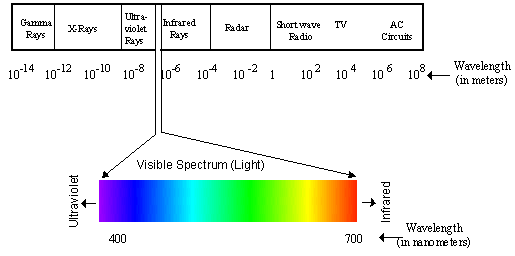
Higher invertebrates and virtually all vertebrates have complex, image-forming eyes, and we will "focus" on the refracting eye found in the octopus and in all vertebrates. Arthropods have compound eyes, which have greater depth of focus than refracting eyes, but which sacrifice resolving power or acuity. Our eyes, like those of many animals, detect a just narrow range of all the wavelengths of electromagnetic radiation, that between 380 and 760 nanometers. This range of light is called the visible spectrum. Figure 1 shows how the visible spectrum fits into the entire electromagnetic spectrum.
 Figure 1. The electromagnetic spectrum and the visible spectrum.
2. The eyeball is an optical device for focusing light
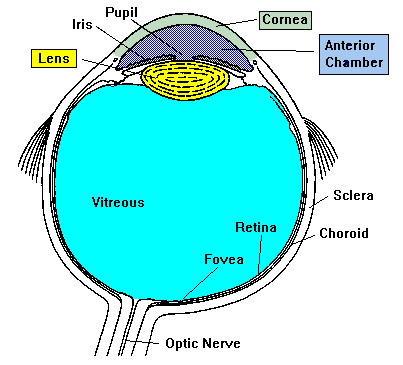
The mammalian eyeball (Figure 2) is an organ that focuses a visual scene onto a sheet of specialized neural tissue, the retina, which lines the back of the eye. Light from a scene passes through the cornea, pupil, and lens on its way to the retina. The cornea and lens focus light from objects onto photoreceptors, which absorb and then convert it into electrical signals that carry information to the brain. Two pockets of transparent fluid nourish eye tissues and maintain constant eye shape: these are the aqueous and vitreous humors, through which the light also passes. The lens projects an inverted image onto the retina in the same way a camera lens projects an inverted image onto film; the brain adjusts this inversion so we see the world in its correct orientation. To control the images that fall upon our retinas, we can either turn our heads or turn our eyes independently of our heads by contracting the extraocular muscles, six bands of muscles that attach to the tough outside covering, or sclera, of the eyeball and are innervated by cranial nerves. See Table 1 for a brief list of eyeball components and their functions.
The cornea and lens bend or refract light rays as they enter the eye, in order to focus images on the retina. The eye can change the extent to which rays are bent and thus can focus images of objects that are various distances from the observer, by varying the curvature of the lens. The ciliary muscle accomplishes this by contracting to lessen tension on the lens and allowing it to round up so it can bend light rays more, or relaxing for the opposite effect. This ciliary muscle is smooth or non-voluntary muscle-you cannot "decide" to contract or relax it as you do the skeletal muscle in a finger or facial muscle.
 Figure 2. The mammalian eyeball.
3. Refractive errors in the eye cause focusing problems
Refractive errors occur when the bending of light rays by the cornea and lens does not focus the image correctly onto the retina. An eyeball that is too long or too short for the optics of the cornea and lens or an irregularly shaped cornea can cause refractive errors, which include myopia (near-sightedness), hyperopia (far-sightedness), and astigmatism. Myopia results either when the eyeball is too long or when the cornea is curved too much, and the focused image falls in front of the retina. Hyperopia is the opposite, with the image falling behind the retina. Astigmatism results from a cornea that is not spherical. Fortunately, most refractive errors can be corrected with prescription lenses.
| TABLE 1. PARTS OF THE EYE |
| STRUCTURE |
FUNCTION |
| Aqueous humor |
clear watery fluid found in the anterior chamber of the eye; maintains pressure and nourishes the cornea and lens |
| Vitreous humor |
clear, jelly-like fluid found in the back portion of the eye: maintains shape of the eye and attaches to the retina |
| Blind spot |
small area of the retina where the optic nerve leaves the eye: any image falling here will not be seen |
| Ciliary muscles |
involuntary muscles that change the lens shape to allow focusing images of objects at different distances |
| Cornea |
transparent tissue covering the front of the eye: does not have blood vessels; does have nerves |
| Cones |
photoreceptors responsive to color and in bright conditions; used for fine detail |
| Rods |
photoreceptors responsive in low light conditions; not useful for fine detail |
| Fovea |
central part of the macula that provides sharpest vision; contains only cones |
| Iris |
circular band of muscles that controls the size of the pupil. The pigmentation of the iris gives "color" to the eye. Blue eyes have the least amount of pigment; brown eyes have the most |
| Lens |
transparent tissue that bends light passing through the eye: to focus light, the lens can change shape |
| Macula |
small central area of the retina that provides vision for fine work and reading |
| Optic nerve |
bundle of over one million axons from ganglion cells that carry visual signals from the eye to the brain |
| Pupil |
hole in the center of the eye where light passes through |
| Choroid |
Thin tissue layer containing blood vessels, sandwiched between the sclera and retina; also, because of the high melanocytes content, the choroid acts as a light-absorbing layer. |
| Retina |
layer of tissue on the back portion of the eye that contains cells responsive to light (photoreceptors) |
| Sclera |
tough, white outer covering of the eyeball; extraocular muscles attach here to move the eye |
|
Comments (0)
You don't have permission to comment on this page.